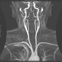
CT imaging uses X-rays in conjunction with computing algorithms to image the body. In CT, an X-ray generating tube opposite an X-ray detector (or detectors) in a ring shaped apparatus rotate around a patient producing a computer generated cross-sectional image. CT is acquired in the axial plane, while coronal and sagittal images can be rendered by computer reconstruction.
Ultrasonography uses high-frequency sound waves to visualize soft tissue structures in the body in real time. No ionizing radiation is involved, but the quality of the images obtained using ultrasound is highly dependent on the skill of the ultrasonographer performing the exam and patient body habitus.
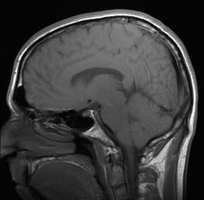
MRI radio signals are collected by small antennae, called coils, placed near the area of interest. An advantage of MRI is its ability to produce images in uses strong magnetic fields to align atomic nuclei within body tissues, then uses a radio signal to disturb the axis of rotation of these nuclei and observes the radio frequencyaxial, coronal, sagittal and multiple oblique planes with equal ease. MRI scans give the best soft tissue contrast of all the imaging modalities. With advances in scanning speed and spatial resolution, and improvements in computer 3D algorithms and hardware, MRI has become an important tool in musculoskeletal radiology and neuroradiology.
Mammography is the process of using low-energy X-rays (usually around 30 kVp) to examine the human breast and is used as a diagnostic and a screening tool. The goal of mammography is the early detection of breast cancer, typically through detection of characteristic masses and/or microcalcifications.
Department Animated Video
- Computer Tomography
- Magnetic Resonance Imaging